
Figure 6.
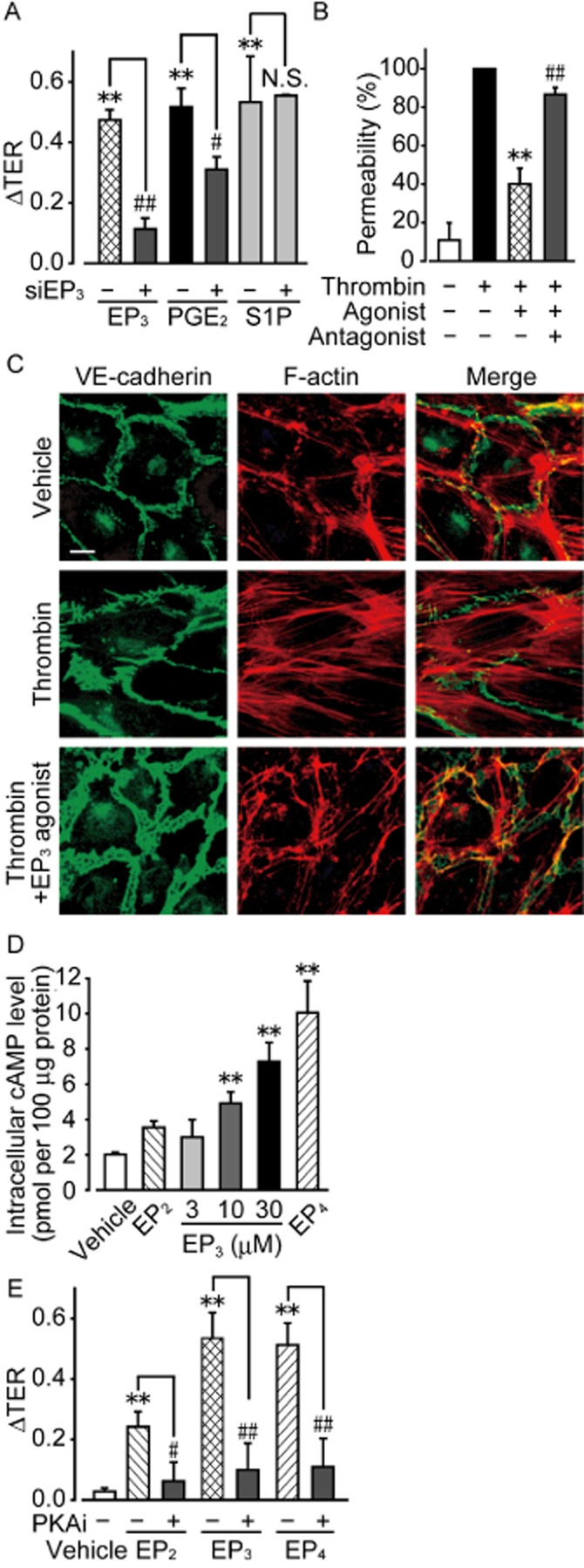
EP3 receptor agonism enhances the endothelial barrier in vitro. (A) HUVECs were transfected with either control or EP3 receptor siRNA. The maximum increase in TER induced by EP3 receptor agonist, PGE2, or sphingosine-1-phosphate (S1P) was quantified (6 ≤ n ≤ 9). (B) FITC-dextran permeability assay (5 ≤ n ≤ 6). (C) Typical pictures of immunostaining of VE-cadherin (left panels, green) and F-actin (middle panels, red) after thrombin stimulation with and without EP3 receptor agonist. Right panels show merged pictures of VE-cadherin, F-actin, and DAPI (blue) staining. Scale bar is 10 μm. (D) Measurement of intracellular cAMP level in HUVECs after stimulation of EP3 receptors (n = 4). (E) Effects of PKAi on EP3 receptor-induced increases in TER (n = 6). **P < 0.01 significantly different from the results of the vehicle treatment. #P < 0.05, ##P < 0.01 significantly different from the results in HUVECs infected with control siRNA (A) or after stimulation by EP3 receptor agonist without any pretreatment (B, D). Data are presented as means ± SEM.