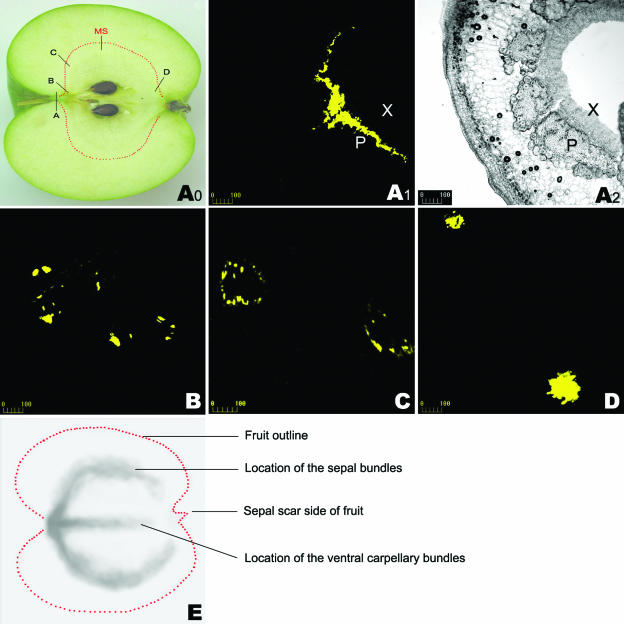
Figure 4.
CF CLSM imaging shown by serial transverse sections throughout whole fruit and 14C-autoradiography at the late developmental stage. CF reached the phloem of the fruit flesh 4 h after being supplied to the fruit pedicel. The treated fruit was harvested 72 h after CF supply. A0, Longitudinal section of apple fruit showing the sampling locations for A1, A2, B, C, and D. A1, CLSM imaging in the transverse section of a portion of the CF-loaded pedicel close to the fruit flesh (see A0), showing that CF was restricted to the phloem zone of the vascular bundle. The corresponding bright-field image is shown in A2. B–D, CLSM imaging of CF in the transverse sections at the different points in fruit (see A0 for the locations of B, C, and D). E, 14C-unloading is apparent along the sepal bundles and the ventral carpellary bundles in fruit. MS in the Figure A0, main sampling site for CLSM imaging at different stages of fruit development, as indicated in Figure 1A; P, phloem; X, xylem. Bars = 100 μm (for Fig. 4, A1–D).

