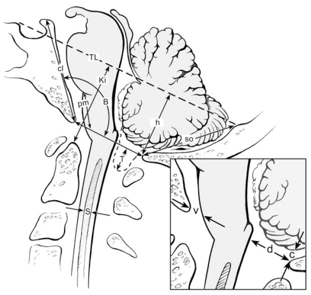
Figure 2.
Measurements were taken from sagittal slice T1-weighted MR images as indicated in this drawing and included tonsillar herniation (T); posterior fossa height (h); Klaus index (Ki); pontomedullary height (pm); supraocciput (so); clivus (cl); Boogaard angle (B); cerebellum to opisthion (c); and ventral subarachnoid space (v); dorsal subarachnoid space (d); syrinx diameter (s); and Twining line (TL; tuberculum sellae to internal occipital protuberance).

