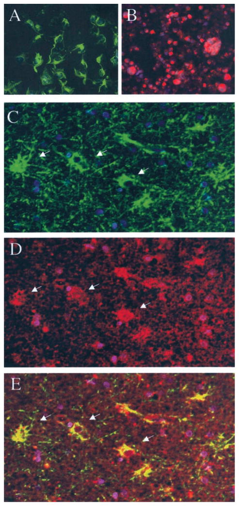
Figure 4.
Co-localization of anti-human herpesvirus 6 (anti-HHV-6) gp116/54/64 reactivity in glial fibrillary acidic protein (GFAP)–reactive cells using double-immunofluorescence assay on formalin-fixed paraffin-embedded brain tissue. Positive staining for GFAP (green) but not for HHV-6 gp116/54/64 (red) of astrocytic cell line U251 was visualized with both fluorescein isothiocyanate and rhodamine filters (A). Positive staining for HHV-6 gp116/54/64 but not for GFAP of HHV-6B (strain Z29)–infected SupT-1 cell line (B) was seen after simultaneous incubation with both sets of antibodies. Lateral temporal lobe of Patient 2 shows presence of GFAP-reactive cells (green) (C, arrows) and of HHV-6 gp116/54/64-positive cells (red; D, arrows) incubated with both anti-GFAP and anti-gp116/54/64 primary antibodies. Juxtaposition of the combined images shows co-localization of GFAP and HHV-6 gp116 reactivity (yellow) (E, arrows) (20× magnification). All fields were counterstained with 4,6-diamidino-2-phenylindole (blue) to visualize nuclei.

