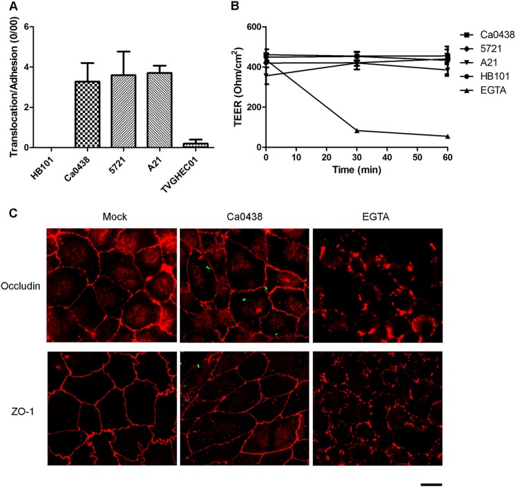
FIG 3.
Translocation of K. pneumoniae cells across polarized intestinal cell monolayers. (A) Translocation assays using human Caco-2 cell monolayers. The translocation rate was expressed after normalization for adhesion. Each bar indicates the mean ± SEM. For Ca0438 and HB101, 5721 and HB101, A21 and HB101, Ca0438 and TVGHEC01, 5721 and TVGHEC01, and A21 and TVGHEC01, all P values are <0.05. (B) TEER measurements of Caco-2 cell monolayers during K. pneumoniae infection at the time points indicated. For comparison, cells were incubated with HB101 or EGTA (10 mM), a chemical that disrupted cell TJs. Three independent experiments were conducted. Data are presented as means ± SEM. (C) Representative immunofluorescence micrographs illustrating cell TJs during K. pneumoniae Ca0438 infection. Differentiated Caco-2 cells were infected by K. pneumoniae Ca0438 cells carrying pCRII-TOPO::GFP (green) or were incubated with EGTA for 1 h. Two major TJ proteins, ZO-1 and occludin, were immunostained with rabbit anti-ZO-1 (upper graphs) or anti-occludin (lower graphs) and the secondary antibody Alexa Fluor 594 anti-rabbit IgG (red). Scale bar, 10 μm.

