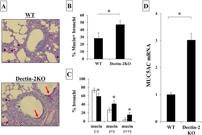
FIG 4.
Mucin production after infection with C. neoformans. WT and Dectin-2KO mice were infected intratracheally with C. neoformans. (A) Sections of the lungs on day 14 postinfection were stained with PAS and observed under a light microscope at ×200 magnification. Arrows indicate the mucin secreted by bronchoepithelial cells. Representative pictures from the five to six mice are shown. The proportion of mucin-producing bronchi (B) and the classification of mucin-producing bronchi (C) were determined. Each bar represents the mean ± SD of results for five to six mice. Experiments were repeated twice, with similar results. *, P < 0.05. (D) MUC5AC mRNA expression in the lungs was measured on day 7 after infection. Each bar represents the mean ± SD of results for three to four mice. Experiments were repeated twice, with similar results. *, P < 0.05.

