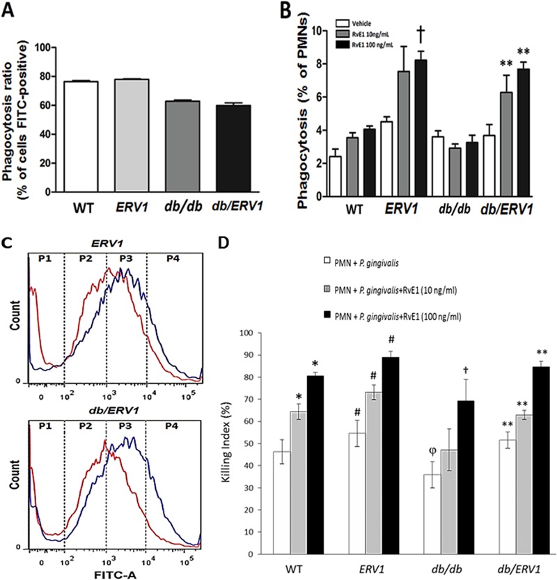
FIG 2.
RvE1 increases P. gingivalis phagocytosis by neutrophils overexpressing ERV1. P. gingivalis phagocytosis by neutrophils was quantified by flow cytometry as the percentage of neutrophils containing bacteria (FITC positive) and the number of bacteria per neutrophil. (A) Percentages of FITC-positive neutrophils. Diabetic mice exhibit significantly fewer phagocytizing neutrophils (n = 9). (B) Percentages of FITC-positive neutrophils with more than 10,000 events (†, P < 0.05 compared to ERV1 with vehicle; **, P <0.01 compared to db/ERV1 with vehicle. (C) Distribution of bacteria per neutrophil by quartile. P4 = >10,000 events per cell. Note the shift to more bacteria per neutrophil with RvE1 treatment (red, no treatment; blue, 10 ng/ml RvE1; n = 8 nondiabetic and 6 diabetic mice). (D) Neutrophil bactericidal activity is enhanced by RvE1. With a CFU killing assay for P. gingivalis, the actions of two doses of RvE1 (10 and 100 ng/ml) were assessed in WT, ERV1, db/db, and db/ERV1 mice. Differences within and between groups were determined by ANOVA with Bonferroni corrections for multiple comparisons. Between-group comparisons revealed that bactericidal activity was lower in db/db mice than in WT animals (or all other strains) (φ, P < 0.05). Treatment of db/db neutrophils with RvE1 restored the killing response to the level of untreated WT mice at 10 ng/ml, and the effect was significantly greater at 100 ng/ml (†, P < 0.05). Overexpression of ERV1 in diabetic db/ERV1 mice completely normalized the killing response (**, P < 0.05). It is interesting that the db/db killing response was significantly lower than that of the WT at both doses of RvE1 and that this response deficiency was eliminated by the overexpression of ERV1. *, P < 0.05 compared to PMNs plus P. gingivalis; #, P <0.05 compared to the WT under all conditions (n = 4).

