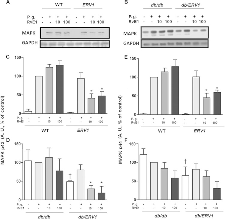
FIG 4.
P. gingivalis (P.g.) induces phosphorylation of MAPK that is reversed by RvE1. (A, B) Representative Western blot images quantified in panels C to F. (C, D) Densitometric quantification of ERK phosphorylation (p42). (E, F) Densitometric quantification of MAPK phosphorylation (p44). A. U., arbitrary units; *, P < 0.05 compared to ERV1 plus P. gingivalis plus vehicle; †, P < 0.05 compared to db/db without P. gingivalis plus vehicle; *, P < 0.05 compared to db/db plus P. gingivalis plus vehicle; (n = 4).

