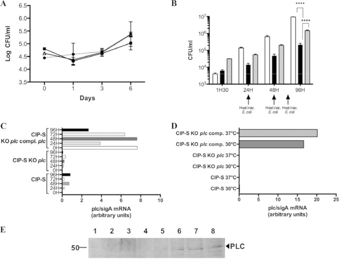
FIG 3.
Survival of plc M. abscessus mutant in eukaryotic cells. (A) Growth of mycobacterial strains within BMDMs recorded by CFU evaluation after 1, 3, and 6 days of coculture. The CIP-S (circles), CIP-S plc KO (squares), and CIP-S plc KO plc-complemented (triangles) strains were used. (B) Growth of mycobacterial strains within amoebae recorded by CFU evaluation after 1.5 h and 1, 2, and 4 days of coculture. CIP-S (white bars), CIP-S plc KO (black bars), and CIP-S plc KO plc-complemented (gray bars) strains were used. Experiments were repeated five times in triplicate at different times for both panels A and B (***, P < 0.001). (C) mRNA plc/sigA ratio (in arbitrary units) for the CIP-S, CIP-S plc KO, and CIP-S plc KO plc-complemented strains cocultivated with macrophages for 5 days. (D) mRNA plc/sigA ratio (in arbitrary units) for the CIP-S, CIP-S plc KO, and CIP-S plc KO plc-complemented strains cultivated in rich medium (7H9) at 30°C or 37°C. The results are representative of two independent experiments (C and D). (E) Western blot analysis of PLC expression during coculture of mycobacterial strains with A. castellanii. Lane 1, total extract (30 μg) of CIP-S cultivated in 7H9 medium; lane 2, total extract (30 μg) of amoebae cultivated for 96 h in PAS buffer in the absence of mycobacteria; lanes 3 to 7, total extract (30 μg) of amoebae cocultivated for 3 h (lane 3), 24 h (lane 4), 48 h (lane 5), 72 h (lane 6), or 96 h (lane 7) in PAS buffer in the presence of CIP-S; lane 8, total extract (30 μg) of amoebae cocultivated for 48 h in PAS buffer in the presence of the CIP-S plc KO plc-complemented strain. This picture is representative of three independent experiments.

