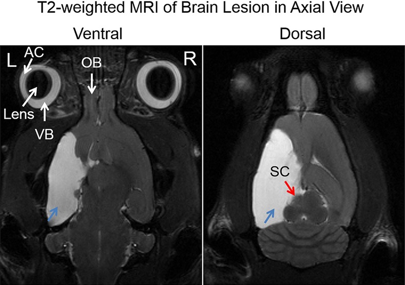
Figure 1.

Axial T2-weighted MRI of the adult rat brain, showing the extent of cystic lesions in the left cortical hemisphere (blue arrows) at 1 year after unilateral neonatal hypoxic-ischemic injury at postnatal day 7. Note also the smaller left superior colliculus (red arrow) compared with the right hemisphere. AC, anterior chamber; VB, vitreous body; OB, olfactory bulb.
