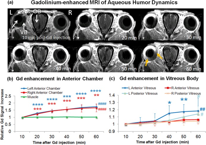
Figure 2.
(a) Dynamic T1-weighted MRI of aqueous humor dynamics at 10 to 60 minutes after intraperitoneal injection of Gd contrast agent, showing rapid Gd enhancement in the AC of both eyes via the highly permeable blood-aqueous barrier. In the ipsilesional left eye, there was a slow, gradual increase in Gd enhancement in the anterior portion of the VB (yellow arrows). Such enhancement was not apparent in the contralesional right anterior vitreous body, suggestive of the compromise of aqueous-vitreous or blood-ocular barrier in the ipsilesional eye. Ln, lens. (b, c) Time profiles of T1-weighted signal intensities in the anterior chamber and the nearby muscle (b), and in the anterior and posterior VB (C) relative to the first time point after systemic Gd administration. Gd signals continuously enhanced in the anterior chamber of both eyes at similar rates within the 1-hour experimental period (ANOVA, ####P < 0.0001; post hoc Dunnett's tests with first time point, **P < 0.01; ***P < 0.001; ****P < 0.0001). Significant Gd enhancement also was observed at later time points in the ipsilesional left vitreous but not the contralesional right vitreous (ANOVA, #P < 0.05; ##P < 0.01; post hoc Dunnett's tests with first time point, **P < 0.01; *P < 0.05; ***P < 0.001). The anterior portion of the left vitreous appeared to enhance earlier at approximately 40 minutes post Gd administration than the posterior portion of the left vitreous at 60 minutes post GD administration, suggestive of the compromise of aqueous-vitreous or blood-ocular leakage leading to the Gd flow from the anterior to posterior vitreous. No change in signal intensity was observed in the muscle, or in the contralesional right vitreous (ANOVA, P > 0.05).

