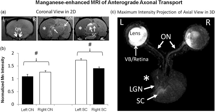
Figure 4.

Manganese-enhanced MRI of anterograde axonal transport along the visual pathways at 1 day after binocular intravitreal Mn2+ injection in 2D multislice coronal view (a) and 3-dimensional maximum-intensity-projected axial view (c). Qualitatively, more Mn enhancement was observed in left VB, left retina, right optic nerve (ON), left superior colliculus (SC), and left lateral geniculate nucleus (LGN) as compared with the opposite hemispheres. The ipsilesional LGN also appeared to be slightly displaced by the cystic lesion in the cortex. Quantitative analysis in (b) showed 14% lower Mn enhancement in the ipsilesional left ON than the right contralesional ON (two-tailed paired t-test, #P < 0.05), whereas the left ipsilesional SC possessed 23% stronger Mn enhancement than the right contralesional SC (two-tailed paired t-test, #P < 0.05). Black bars refer to visual pathway projected from the ipsilesional eye to ipsilesional ON and contralesional SC. White bars refer to visual pathway projected from the contralesional eye to the contralesional ON and ipsilesional SC. Asterisks indicate hypointense brain lesion in cortical regions.
