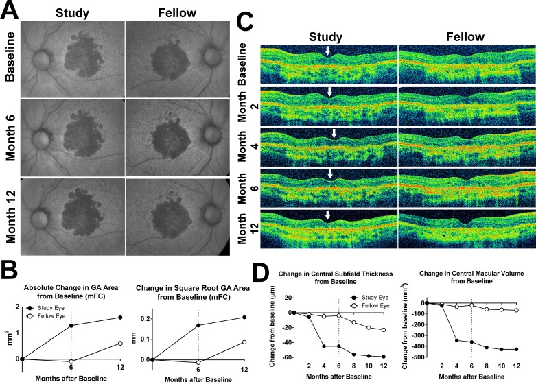
Figure 2.
Fundus changes in participant 5 during study treatment and follow-up. (A) mFC FAF images demonstrated bilateral central GA lesions at baseline that gradually expanded in both study and fellow eye in a manner not inconsistent with the natural history of GA. (B) Absolute (left) and square root transform (right) increase in GA area in the study and fellow eye at month 6 and month 12 were plotted, demonstrating a greater increase in GA area in the study eye at month 12 compared with that in the fellow eye. (C) Horizontal aligned B-scans through the center of the macula from baseline to month 12 were obtained. Longitudinal analysis demonstrated noticeable thinning of the central macula beginning at month 4 in the study eye (loci indicated by arrows). Investigational product administration was suspended for the month 6 visit and withheld thereafter. (D) Change in central retinal thickness (left) and central macular volume (right) from baseline to month 12 in the study and fellow eyes documented a rapid decline over the first 4 months in the study eye, which stabilized following drug suspension at month 6 (dashed line), while measures in the fellow eye remained comparatively stable over time.

