
Figure 1. miR-9 was frequently up-regulated in primary ESCC cases and cell lines.
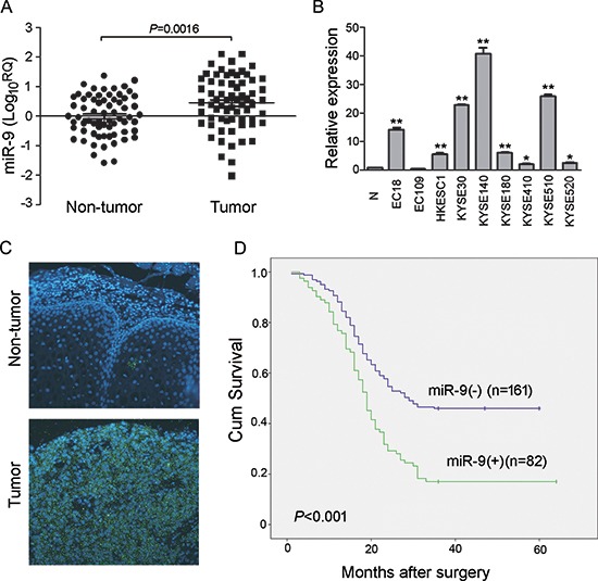
(A) qRT-PCR shows that miR-9 was frequently up-regulated in 67 primary ESCC tissues compared with their adjacent non-tumor tissues. (P = 0.0016, independent t test). Expression of miR-9 was shown in log10 scale and normalized to U6. (B) Up-regulation of miR-9 was detected in all tested ESCC cell lines except EC109 compared with pool of non-tumor tissues (N). U6 was set as an endogenous control. *P < 0.05; **P < 0.001. (C) Representative, of miR-9 expression (green signals) in a pair of ESCC tumor tissue and corresponding non-tumor tissue detected by MISH. Nuclei were conterstained by DAPI (blue color). Original magnification, 40 × objective. (D) Kaplan-Meier analysis indicates up-regulation of miR-9 was significantly associated with poorer overall survival rates of ESCC patients (P < 0.001).