
Figure 2. Tumorigenic effect of doxorubicin and tunicamycin on cell surface GRP78 negative cell lines.
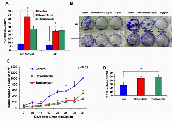
(A) The 4T1 breast cancer mouse cell line expressed a low percent of cell surface GRP78 similar to MDAMB468. Doxorubicin and tunicamycin induced a significant increase in cell surface GRP78 (*p < 0.001). (B) Colony formation by MDAMB468 and 4T1 TNBC cells treated with doxorubicin and tunicamycin was inhibited significantly (*p < 0.001). (C) 10-week-old Balb/C nude mice were inoculated subcutaneously in the right flank with 1 × 10⁶ 4T1 cells in 100 μL PBS or with 4T1 pre-incubated with 0.1 μg/ml doxorubicin or with 10 μg/ml tunicamicin (10 mice per group). Mice from the same group uniformly developed relatively small tumors after doxorubicin or tunicamycin treatment compared to non treated mice cells (p < 0.02). (D) 4T1 cells extracted from mice xenografts, 31 days after tumor inoculation, showed significant increased cell surface GRP78 pre-incubated with doxorubicin (0.1 μg/ml) or tunicamycin (10 μg/ml) (*p < 0.004).