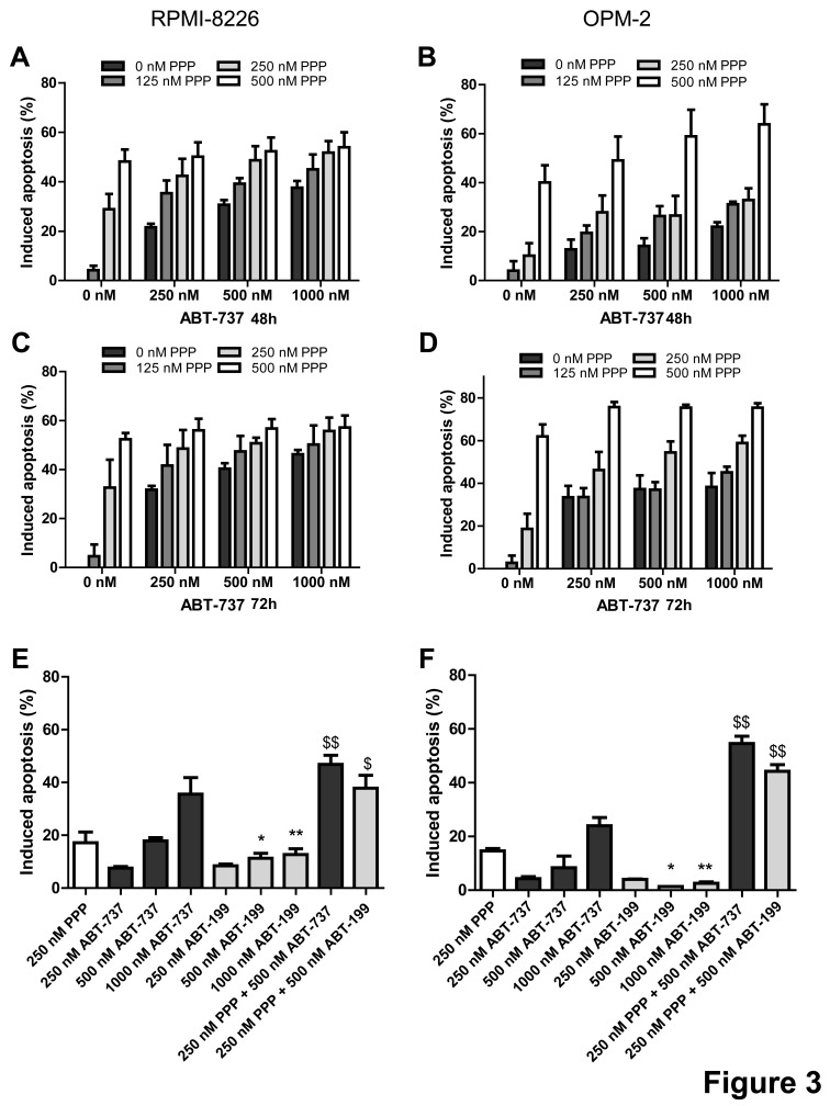
Figure 3. PPP potentiates ABT-737 and ABT-199 mediated apoptosis.
A-D: PPP increased ABT-737 mediated apoptosis. RPMI-8226 (A, C) and OPM-2 (B, D) cells were treated with 0 nM (black bars), 125 nM (dark grey), 250 nM (light grey) or 500 nM (white) PPP either alone or in combination with indicated concentrations of ABT-737 for 48h (A, B) and 72h (C, D). P-values and combination indexes after 48h are shown in Table 1. E-F: PPP increased ABT-199 mediated apoptosis. RPMI-8226 (E) and OPM-2 (F) cells were treated with 250 nM PPP either alone or in combination with indicated concentrations of ABT-737 or ABT-199 for 48h. Effect on apoptosis was determined by an AnnexinV-FITC/7′AAD staining followed by FACS analysis. Results are expressed as the percentage induced apoptosis compared to untreated cells. Columns and error bars are the mean ± SD from at least 3 individual experiments. * and ** indicate p-values of respectively ≤0.05 and ≤0.01 comparing ABT-737 with ABT-199, while $ and $$ indicate p-values of respectively ≤0.05 and ≤0.01 compared to both single agents.

