
Figure 5. Disorganization of cytoskeleton in carcinoma A431 cells caused by VACV strains in vitro and in vivo.
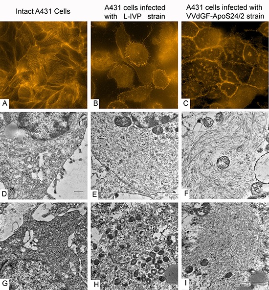
The upper row shows actin filaments visualized in fluorescent microscope with TRITC-falloidin in: (A) intact cell culture, (B) L-IVP strain infected cells, and (C) VVdGF-ApoS24/2 strain infected cells. The middle row shows the same cells visualized in electron microscope: (D) intact cell, (E) infected with the L-IVP strain, and (F) VVdGF-ApoS24/2 strain, clumps of actin are seen. Photos A-F were obtained from A431 cells in vitro, 48 h after the infection with VACV. The bottom row shows electron microscopic images of the A431 cells in vivo: (G) a cell of saline-treated tumor, (H) a tumor cell containing debris and viral particles, 36 days after injection of the L-IVP strain, (I) a tumor cell containing an accumulation of filaments, 36 days after the injection of the VVdGF-ApoS24/2 strain.