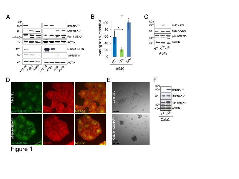
Figure 1. hMENA11a defines an epithelial phenotype and is expressed alternatively to hMENAΔv6 isoform in lung cancer cell lines.
The isoforms have opposite and antagonistic roles in lung cancer cell invasion and affect cell morphology in 2D and 3D cultured cells. (A) WB analysis of lysates of lung tumor cell lines with hMENA isoform specific and pan-hMENA [which recognizes all hMENA isoforms, with apparent molecular weights of 90kDa, hMENA11a, 88kDa, hMENA and 80kDa, hMENAΔv6 (arrow)] and E-CADHERIN and VIMENTIN antibodies, indicating a strong correlation between hMENA11aand E-CADHERIN expression. (B) Matrigel invasion assays of A549 cells transfected with the empty vector (EV), with hMENA11a (11A), or with hMENAΔv6 (Δv6). The invasive ability was measured using Matrigel coated transwell filters towards a serum gradient. The assay was repeated three times and performed in triplicate each time. * Significantly different as determined by Student t tests p=0.027; **p=0.004. (C) WB analysis of A549 cells transfected with the empty vector, with hMENA11a, or with hMENAΔv6, using hMENA isoform-specific Abs or pan-hMENA Ab. (D) Immunofluorescence analysis of A549 cells transfected with the empty vector or with hMENA11a using a pan-hMENA mAb, indicating a colocalization of hMENA isoforms (green) with phalloidin stained actin filaments (red). Cells were imaged using immunofluorescence microscopy DMIRE2 (Leica Microsystems) and processed using FW4000 Software. Magnification: 63X. Scale Bar: 30 μm. (E) Representative phase-contrast images of Calu1 cells transfected with the empty vector (EV) or hMENA11a (11A) and grown for 72h in 3D lrECM. Magnification: 20X. Scale Bar: 100μm. (F) WB analysis of Calu1 cells transfected with the empty vector or with hMENA11a, using hMENA isoform-specific Abs or pan-hMENA Ab.

