
Figure 5. Noxa is necessary for MEK/ERK-driven autophagy in melanoma cells.
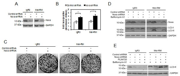
(A) Whole cell lysates from IgR3 and Mel-RM cells stably transduced with the control or Noxa shRNA were subjected to western blot analysis. Data shown are representative of three individual experiments. (B) IgR3 and Mel-RM cells stably transduced with the control shRNA or Noxa shRNA were subjected to cell proliferation assays using the BrdU incorporation method. Data shown are representative of three individual experiments. (n=3, mean ± S.E.M.). *P < 0.05, Student's t-test. (C) IgR3 and Mel-RM cells stably transduced with the control or Noxa shRNA were subjected to clonogenic assays. Data shown are representative of three individual experiments. Scale bar, 1cm. (D) IgR3 and Mel-RM cells stably transduced with the control or Noxa shRNA were treated with or without bafilomycin A1 (100nM) for 2 hours. Whole-cell lysates were subjected to western blot analysis. Data shown are representative of three individual experiments. (E) IgR3 and Mel-RM cells stably transduced with the control or Noxa shRNA were treated with PLX4720 (3μM) or U0126 (20μM) for 24 hours in the presence of bafilomycin A1 (100nM). Whole-cell lysates were subjected to western blot analysis. Data shown are representative of three individual experiments.