
Fig. 3.
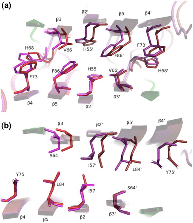
Dimer interface features allowing the shear movement. LC8–nNOS (red/green) is overlaid on apo-LC8 (purple), with alignment based on the lower subunits. The view is along the 2-fold symmetry axis, parallel with the β- strands, in the layer containing (a) Phe86 and (b) Ile57. Note the rotameric interconversion of Ile57. The fluid shear movement of 2 Å is evident in the upper subunits and is facilitated by close residue packing yet lack of side-chain interdigitation.