
Fig.5.
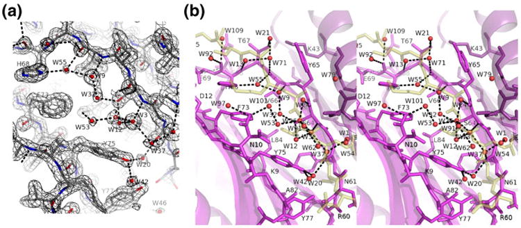
Solvent structure in the apo-LC8 ligand binding groove. (a) 2Fo–Fc density contoured at 1.2σ. Water molecules are shown as red spheres. (b) Ribbon diagram and bound water molecules (red) are shown for apo-LC8 (purple) overlaid with the peptide from LC8–Swa (ghostly yellow). Water 20 (bridging Tyr75 OH and Phe62 O) has an interesting analog in LC8–Swa, where water 1 bridges the same two residues plus Swa Thr293 Oγ and is the only buried water molecule in the structure as well as the most ordered one. An analogous water molecule is missing from LC8–nNOS, due to the different peptide main-chain conformation that leaves insufficient room between the Q−1 and Q+1 residues.