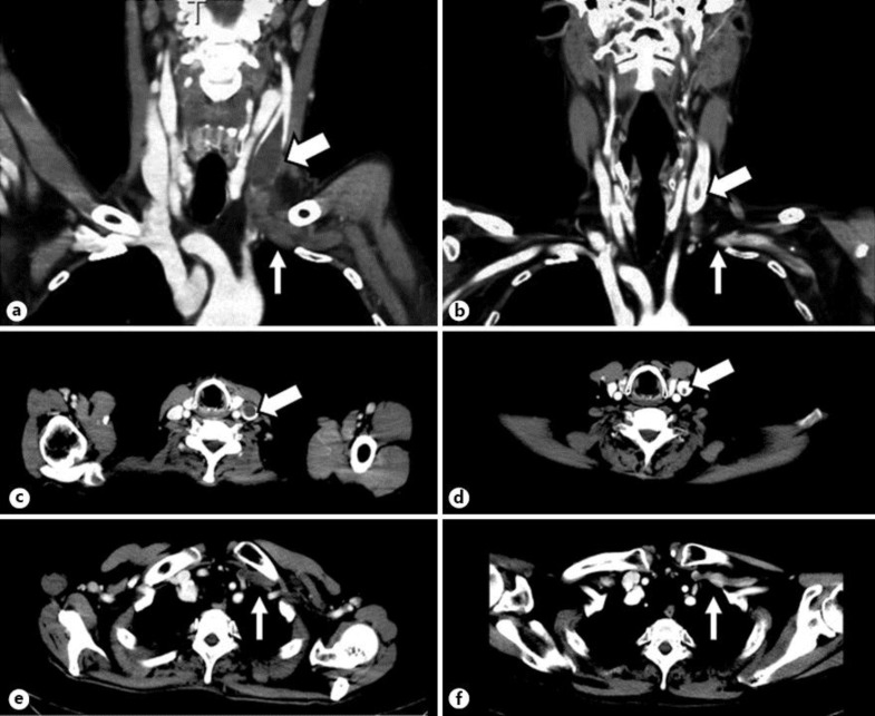
Fig. 1.
Contrast-enhanced CT images on the 54th day of hospitalization (a–c) and 89 days after discharge, i.e. 186 days after admission (d–f). Thrombi were observed in the left internal jugular vein (thick arrows) and the left subclavian vein (thin arrows) as gray masses in coronal (a) and axial views (b, c). 132 days later, the thrombi in the left internal jugular vein (thick arrow) had become smaller in size, and those in the left subclavian vein (thin arrows) had disappeared in coronal (d) and axial views (e, f).

