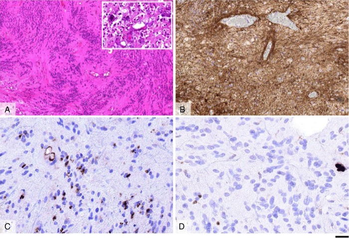
Figure 2:
Morphological appearances of the ependymoma, WHO grade II. Haematoxylin and eosin-stained section (A) shows a moderately cellular tumour with frequent perivascular pseudorosettes and areas of stromal microcalcifications (inlet in A). Immunostaining for GFAP reveals widespread labelling in the tumour cells (B); whereas that for EMA shows frequent perinuclear dot-like labelling and accentuates occasional small ependymal canals (C). The Ki67 proliferation index in the tumour is low (D). Scale bar: 100 µm (A and B), 20 µm (C and D), 60 µm (inlet in A).

