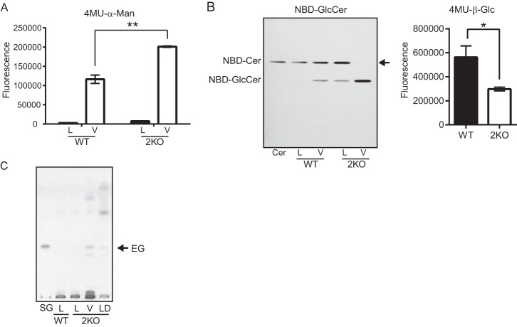
FIGURE 9.
Cellular localization of EG that accumulated in egcrp2-disrupted mutants (2KO) of C. neoformans. A, the α-mannosidase activity of the cell lysate and vacuole fractions. Activity was measured at 37 °C for 1 h using 0.5 μg of protein from each fraction and 20 nmol of 4MU-α-mannoside as a substrate in 100 μl of 50 mm phosphate buffer, pH 6.5. L, lysate; V, vacuole fraction. B, the β-glucosidase activity of the vacuole fraction of WT and 2KO. Activity was measured at 30 °C for 18 h by using 0.25 μg of protein from vacuole fraction and 50 nmol of C6-NBD-GlcCer in 20 μl or 30 nmol of 4MU-β-glucoside as a substrate in 100 μl of 50 mm sodium acetate buffer, pH 5.0. C, TLC showing EG that accumulated in 2KO. Glycolipids were extracted from the lysate and vacuole fractions of WT and 2KO (each 50 μg as protein) and analyzed by TLC using the method described in the legend to Fig. 5A. L, lysate; V, vacuole fraction; LD, lipid droplet fraction. * and **, p < 0.05 and p < 0.0001, respectively. Error bars, S.E.

