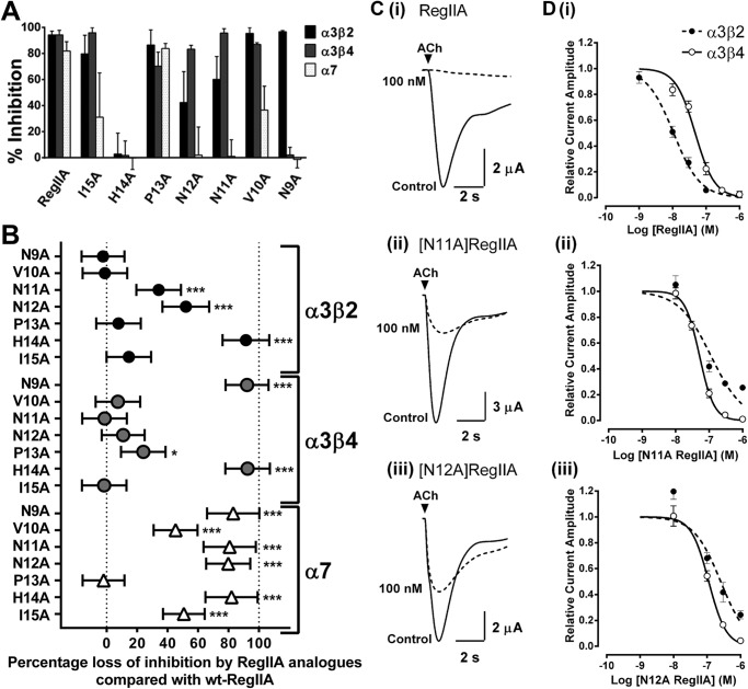
FIGURE 3.
Inhibition of nAChR subtypes expressed in Xenopus oocytes by RegIIA and alanine analogs. A, bar graph of inhibition of nAChR subtypes by RegIIA and its analogs. B, two-way analysis of variance scatter plot illustrating the loss of activity of RegIIA analogs (300 nm) relative to wild-type RegIIA at various nAChR subtypes. [H14A]RegIIA completely lost its activity at α3β2, α3β4, and α7 nAChRs. [N9A]RegIIA was more selective for the α3β2 subtype than RegIIA. [N11A]RegIIA and [N12A]RegIIA selectivity for the α3β4 nAChR subtype significantly improved. All analogs, except [P13A]RegIIA, significantly lost activity at the α7 nAChR subtype. ***, p < 0.001; *, p < 0.05; n = 4–6. C, superimposed traces showing inhibition of α3β2 nAChR-mediated ACh-evoked currents by 100 nm RegIIA (i), [N11A]RegIIA (ii), and [N11A]RegIIA (iii). D, concentration-response curves for RegIIA (i), [N11A]RegIIA (ii) and [N12A]RegIIA (iii) inhibition of the α3β4 (black line, open symbols) and α3β2 nAChR subtypes (dash line, closed symbols). [N11A]RegIIA and [N12A]RegIIA shifted the curve to the right for the α3β2 nAChR subtype, giving an IC50 value of 116 and 278 nm, respectively. All data represents mean ± S.E., n = 4–6.

