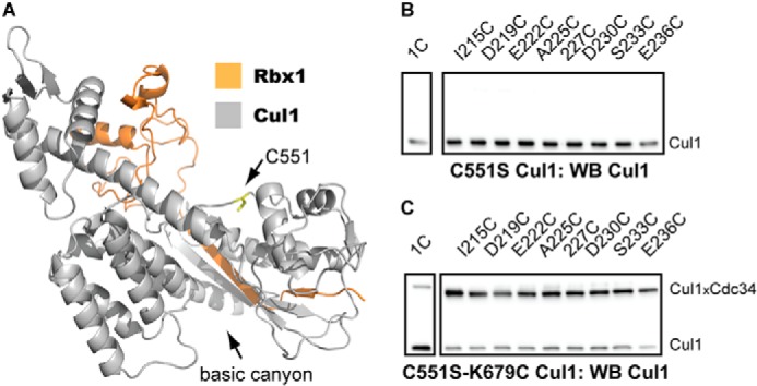
FIGURE 5.

Native cysteine residue Cys-551 in WT Cul1 forms cross-links to the Cdc34 2C proteins. A, ribbon diagram of the C-terminal domain of Cul1 (gray) and Rbx1 (orange). The Cys-551 side chain is shown in a ball-and-stick representation (yellow), and its location is highlighted by the black arrow. The structure is from Protein Data Bank entry 1LDK (16). B, Cul1 Western blot of reactions between either Cdc34 1C or Cdc34 2C proteins and C551S Cul1-Rbx1 in the presence of BMOE. Notice that no Cdc34-Cul1 cross-links are observed. C, Cul1 Western blot of reactions between either Cdc34 1C or Cdc34 2C proteins and C551S/K679C Cul1-Rbx1 in the presence of BMOE. Notice that the introduction of a cysteine residue at Lys-679 in C551S Cul1-Rbx1 restores cross-linking to the Cdc34 2C proteins. The time of incubation for all reactions was 1 min.
