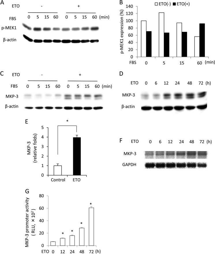
FIGURE 2.
Influence of the senescence-inducing agent ETO on MEK-1 activation and MKP-3 protein level. A–C, levels of phosphorylated MEK-1 and MKP-3 in control and senescent cells. NRK cells were treated with 1 μg/ml ETO for 3 days or left untreated. Then cells were exposed to 5% FBS for the indicated time. Cellular lysates were subjected to Western blot analysis of p-MEK1 (A) and MKP-3 (B). Densitometric quantitation of the level of the phosphorylated MEK-1 in A is shown in C. The result was normalized to zero point control. D–G, changes in MKP-3 during induction of senescence with ETO. Cells were treated with 1 μg/ml ETO for the indicated time and subjected to Western blot analysis of MKP-3. β-Actin was used as a loading control (D). Densitometric quantitation of the level of MKP-3 at the 48-h point following exposure to 1 μg/ml ETO is shown in E. The result was normalized to the control. Data are mean ± S.E. (n = 3). *, p < 0.05 versus control. F, cells were treated with 1 μg/ml ETO for the indicated time and subjected to Northern blot analysis of MKP-3. GAPDH was used as a loading control. G, cells transfected with pGL3B/DUSP6-luc were exposed to 1 μg/ml ETO for the indicated time. Cell lysates were subjected to a luciferase assay. Data are mean ± S.E. (n = 4). RLU, relative light unit. *, p < 0.05 versus zero point control.

