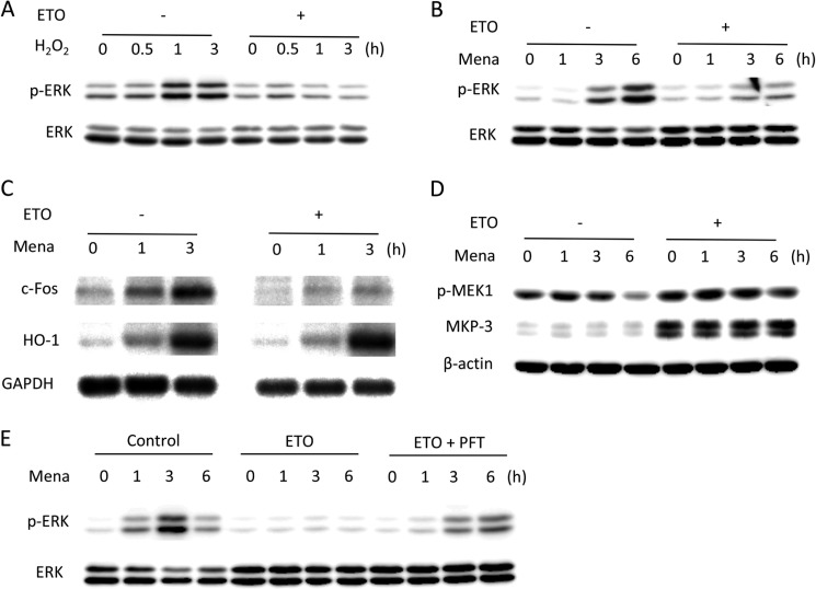
FIGURE 7.
Impaired ERK1/2 activation in oxidant-exposed senescent cells. A–C, ERK1/2 phosphorylation induced by oxidative stress in senescent cells. Control and senescent cells were exposed to 100 μm H2O2 (A) or 50 μm menadione (Mena, B) for the indicated times and subjected to Western blot analysis of p-ERK1/2 and ERK1/2. C, cells were exposed to 50 μm menadione for the indicated time and subjected to Northern blot analysis of c-Fos and Ho-1. GAPDH was used as a loading control. D and E, involvement of p53 in the regulation of ERK1/2 phosphorylation. D, control and senescent cells were treated with 50 μm menadione for the indicated time and subjected to Western blot analysis of p-MEK1 and MKP-3. β-Actin was used as a loading control. E, control cells were treated with 1 μg/ml ETO in the absence or presence of 5 μm PFT for 3 days, and then cells were exposed to 50 μm menadione for the indicated time. Cell lysates were subjected to Western blot analysis of p-ERK1/2 and ERK1/2.

