FIGURE 1.
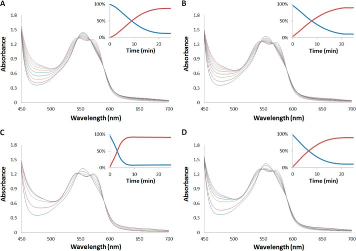
Effect of XOR on nitrite reduction by Hb. Time resolved absorbance spectra after mixing deoxyhemoglobin (100 μm) and nitrite (500 μm) in the presence of sodium dithionite (10 mm) with NADH (1 mm) (t½ = 9 min) (A), NADH (1 mm) and XOR (1 μm) (t½ = 7.8 min) (B), and NADH (1 mm) and XOR (10 μm) (t½ = 3 min) (C), and NADH (1 mm), XOR (10 μm), and allopurinol (100 μm) (t½ = 6.6 min) (D). Insets show the percentage of deoxyHb (blue) and HbNO (red) as a function of time. Spectra were recorded at 37 °C every 128 s for data shown in panels A, C, and D and every 180 s for data shown in panel B. Samples were all prepared in 100 mm HEPES buffer, pH 6.5.

