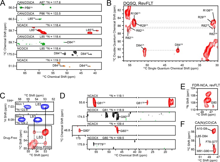
FIGURE 3.
Spectroscopic characterization of the loop connecting TM3 and TM4 in the drug-free state in DMPC-liposomes. A, the backbone resonances for [U-13C,15N]EmrE were assigned with MAS-SSNMR using a backbone walk with CAN(CO)CA (green), NCACX (red/orange), and NCOCX (black) experiments. Note that the orange NCACX (Asp84) was collected with revWIY-labeled EmrE. B, DQSQ experiment with revFLT shows the arginine residues clustered around 55 ppm. Unlike Arg29 and Arg106, Arg82 is split into two distinct peaks, which results in a weaker signal overall. The assignment for Arg82 is also in good agreement with data collected from solution NMR (Fig. 4). C, similarity of chemical shifts in the TPP+-bound form from solution NMR (blue) are directly transferable to spectra collected in the drug-free form with MAS-SSNMR, which provided the assignment for Leu83 in an 800-ms PDSD experiment on [2-13C,15N]Leu EmrE. D, Gln81, Gly80, and Phe79 are visible in NCACX and NCOCX spectra collected at 750 MHz (1H frequency) with [U-13C,15N]EmrE in 3/1 DMPC/DMPG and are in good agreement with data from solution NMR. E, the FDR-NCA with revFLT served as a spectroscopic filter that highlighted residues i + i relative to the reverse labeled amino acids and allowed for the assignment of Gly80 (28). F, a two-dimensional CA(N)(CO)CA with [U-13C,15N]EmrE was used to assign Gly77 and Phe78.

