FIGURE 1.
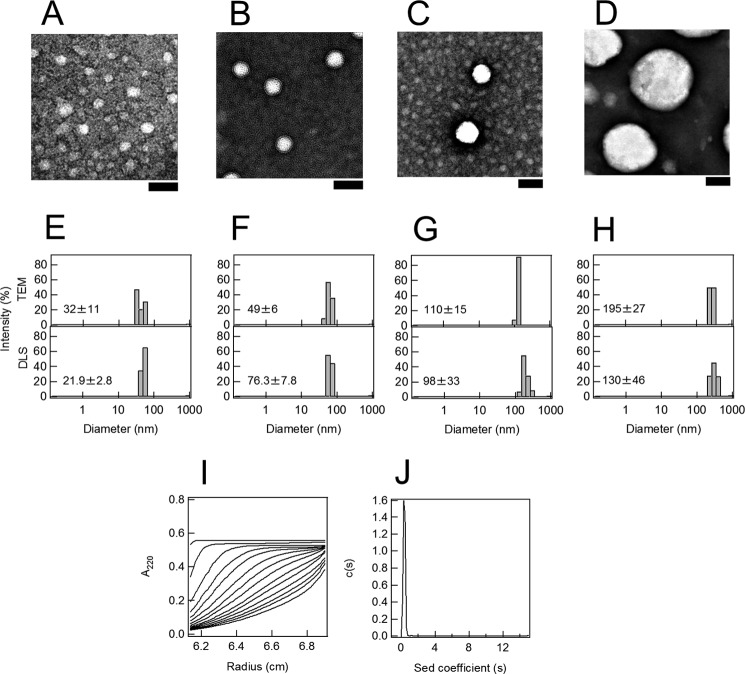
Preparation of liposomes of various sizes and Aβ monomers. A–D, TEM images of liposomes of various diameters: 30 (A), 50 (B), 100 (C), and 200 nm (D). The scale bar represents 100 nm. E–H, distribution of liposome diameters determined by TEM and dynamic light scattering (DLS). The liposome diameters prepared were 30 (E), 50 (F), 100 (G), and 200 (H) nm. I, sedimentation boundary profiles of Aβ monomers. A sedimentation pattern was recorded at 60,000 rpm and 4 °C by monitoring the absorbance at 220 nm, and fitted traces at intervals of 20 min are shown. J, sedimentation coefficient distributions derived from sedimentation boundary profiles of Aβ monomers.

