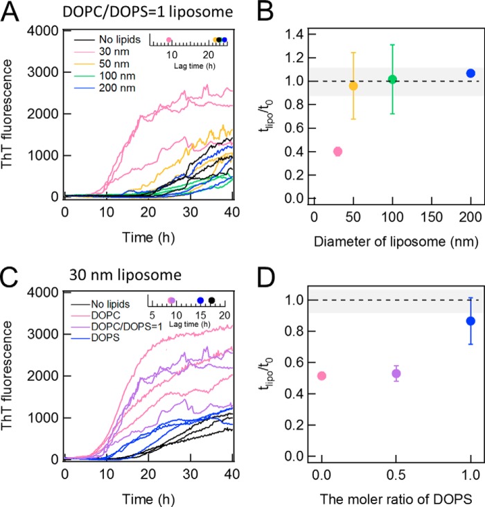
FIGURE 7.
Effects of DOPS on the DOPC-dependent fibrillation of Aβ-(1–40). A and B, the kinetics of Aβ-(1–40) fibrillation in the presence of 50% DOPS liposomes of 30 (pink), 50 (yellow), 100 (green), or 200 (blue) nm in diameter or without lipids (black) (A) and the dependence of relative lag time on the liposome size (B). The inset in A represents the average lag time at various liposome sizes. C and D, the kinetics of Aβ(1–40) in the presence of 100% DOPC (pink), 50% DOPS (purple), and 100% DOPS (blue) liposomes of 30 nm in diameter (C) and the dependence of relative lag time on the DOPC content (D). The inset in C represents the average lag times at different contents of DOPS.

