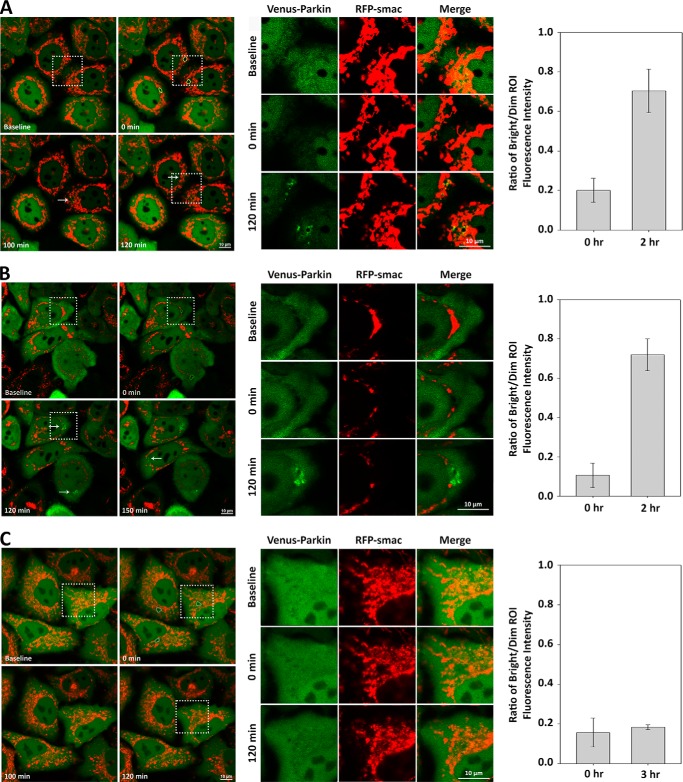
FIGURE 7.
ATP-dependent PINK1 expression mediate Parkin mitochondrial translocation. A–C, inhibition of cellular bioenergetics at different stages verifies dependence on ATP to instigate Parkin mitochondrial translocation. HeLa cells expressing Venus-Parkin-WT and RFP-smac-mts were incubated in either 30 mm 2-DG (A), 10 μm oligomycin (B), or both (C) for 2 h, followed by individual mitochondrial photoirradiation with 405-nm light (indicated by dotted white outlines) and visualized for Parkin accumulation (indicated by arrows) in the zoomed in view of the dotted square regions within the left panels. Parkin aggregation fold difference in fluorescence intensity versus background was quantified by acquiring ratio between bright versus dim ROI.

