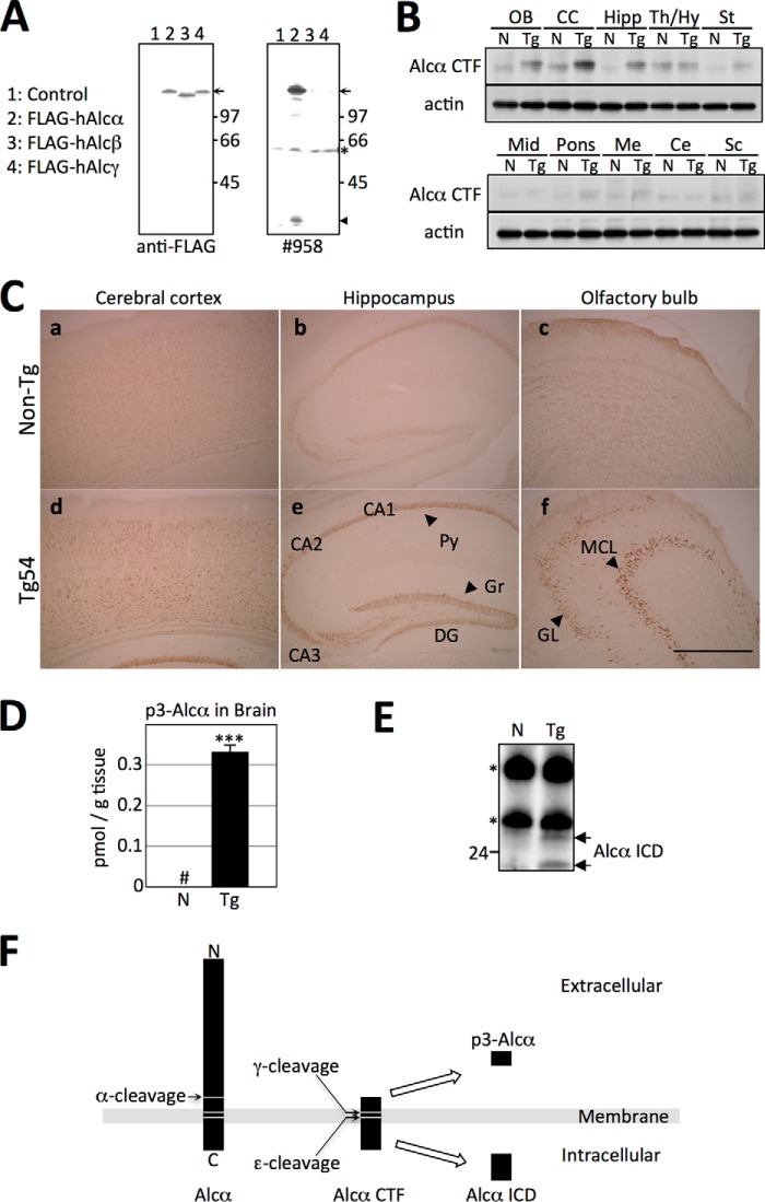
FIGURE 2.
Expression of hAlcα CTF in the brains of Tg54 mice. A, specificity of anti-Alcα carboxyl-terminal domain antibody no. 958. Lysates of HEK293 cells expressing human FLAG-Alcα (lane 2), human FLAG-Alcβ (lane 3), and human FLAG-Alcγ (lane 4) along with plasmid alone (lane 1) were analyzed by immunoblotting with anti-FLAG M2 (left) and anti-Alcα no. 958 (right) antibodies. The arrow indicates FLAG-tagged Alcα, Alcβ, and Alcγ, and the arrowhead indicates Alcα CTF. Numbers indicate protein standards (kDa). The asterisk indicates a nonspecific product. B, expression of hAlcα CTF in the brain regions of Tg54 mice. Expression of hAlcα CTF in brain regions of Tg54 mice was examined by immunoblotting with no. 958 antibody. Brain tissue from 3-month-old Tg54 (Tg) and non-Tg (N) littermates were dissected into the indicated brain regions, and lysates obtained from these sections were analyzed by immunoblotting with no. 958 (top) and anti-β-actin (bottom) antibodies. OB, olfactory bulb; CC, cerebral cortex; Hipp, hippocampus; Th/Hy, thalamus/hypothalamus; St, striatum; Mid, midbrain; Me, medulla; Ce, cerebellum; Sc, spinal cord. C, localization of hAlcα CTF in the brains of Tg54 mice. Localization of hAlcα CTF in brain regions was examined by immunostaining with no. 958 antibody. Sections of the cerebral cortex (a and d), hippocampus (b and e), and olfactory bulb (c and f) were prepared from 3-month-old Tg54 (d–f) and non-Tg (a–c) littermates and immunostained. Py, pyramidal cells; Gr, granule cells; DG, dentate gyrus; MCL, mitral cell layer; GL, granule cell layer. D, quantification of p3-Alcα in the brains of Tg54 mice. The total amount of p3-Alcα in the brains of 6-month-old Tg54 (Tg, filled bar) and non-Tg (N, open bar) littermates were quantified by sELISA. Quantified values are given as the mean ± S.E. (n = 4). ***, p < 0.005, Student's t test. #, below detectable levels. E, detection of hAlcα ICD in the cytosolic fraction of mice brains. Brain lysates prepared from 2-month-old Tg54 (Tg) and non-Tg (N) mice brains were subjected to immunoprecipitation with no. 958 antibody, and the precipitates were detected by immunoblotting with the same antibody. Arrows indicate hAlcα ICD fragments. Numbers on the left side of the panel indicate the molecular mass (kDa). Asterisks indicate IgG heavy and light chains. F, schematic structure of p3-Alcα and hAlcα ICD. Alcα CTF is first cleaved at the ϵ-site by γ-secretase to release Alcα ICD into the cytoplasm. Next, γ-secretase cleavages reach to the γ-site to secrete p3-Alcα into the extracellular milieu (15).

