
Figure 1.
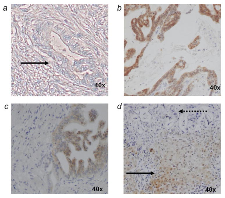
Immunohistochemical analysis of ASS expression in human pancreatic tumors. (a) pancreatic adenocarcinoma lacking ASS expression (solid arrow), (b) pancreatic adenocarcinoma expressing ASS, (c) benign pancreatic ductal cells expressing ASS, (d) metastatic pancreatic adenocarcinoma lacking ASS expression (dotted arrow) with adjacent hepatocytes demonstrating ASS expression (solid arrow). In panels a–c, only neoplastic glandular tissue of pancreatic adenocarcinoma is observed within a dense field of fibroblasts typical of the dense desmoplastic reaction and chronic pancreatitis of pancreatic adenocarcinoma.