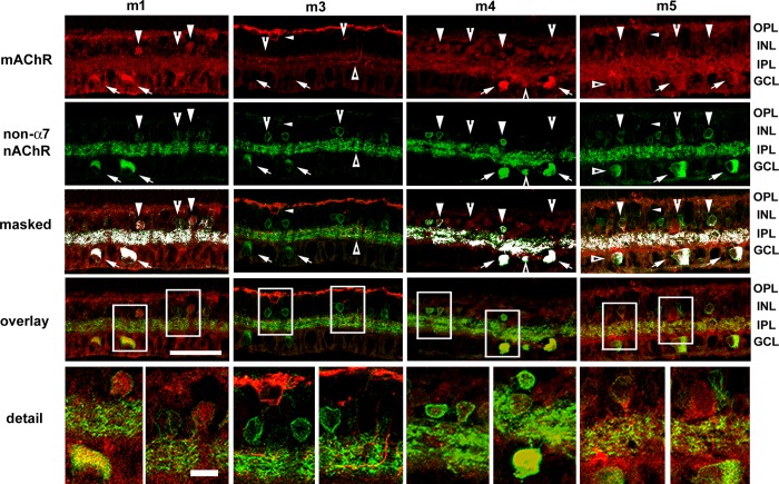
Fig. 1.
Expression patterns of muscarinic ACh receptors (mAChRs) relative to the expression patterns of non-α7 nicotinic AChRs (nAChRs). Confocal image stacks of projections of 5 optical sections (0.5 μm steps) of m1 and m3–m5 mAChRs relative to β2-containing nAChRs. Colocalization was assessed from single confocal optical sections (0.5 μm steps) and masked onto overlay images. Column 1: many ganglion cells (arrows) and subsets of amacrine cells were immunoreactive for both m1 mAChRs and β2-containing nAChRs (arrowheads). Whereas most m1-positive amacrine cells also displayed β2-containing nAChR immunoreactivity (IR), not all amacrine cells that were positive for β2-containing nAChRs were immunoreactive for m1 receptors (notched arrowheads). Whereas both m1 and β2-containing nAChR IR was broadly distributed throughout the inner plexiform layer (IPL), the limited colocalization in the IPL was consistent with the somatic-labeling patterns. Muscarinic m1 IR was present in the outer plexiform layer (OPL), possibly indicating m1 expression by bipolar cells or horizontal cells. Column 2: there was no apparent double-labeling of m3 mAChRs and β2-containing nAChRs. Arrows and notched arrowheads indicate ganglion and amacrine cell somas that displayed β2-containing nAChR IR, whereas small arrowheads indicate bipolar cells that displayed m3 mAChR IR. m3 IR processes were localized to the OPL and to innermost and outermost sublamina (triangles) of the IPL, whereas β2-containing nAChR IR was broadly distributed throughout the IPL. Column 3: m4 muscarinic IR and β2-containing nAChR IR were colocalized almost completely in cell bodies in the ganglion cell layer (GCL; arrows) but were only colocalized to subsets of cell bodies in the inner nuclear layer (INL; arrowheads), whereas other m4-immunoreactive amacrine cell somata did not display β2-containing nAChR IR (notched arrowheads). Double-labeled processes were evident throughout the IPL. Column 4: ganglion cells in the GCL (arrows) and subsets of amacrine cells in the INL (arrowheads) were double labeled for m5 muscarinic and β2-containing nAChRs, and double-labeled processes were clearly visible throughout the IPL. Additional ganglion (triangles) and amacrine cells (notched arrowheads) were immunoreactive for β2-containing nicotinic but not m5 mAChRs. Bipolar cells (small arrowheads) were immunoreactive for m5 mAChRs but not β2-containing nAChRs. Original scale bar, 50 μm. Bottom: boxed areas in detail; original scale bar, 10 μm.

