
Fig. 1.
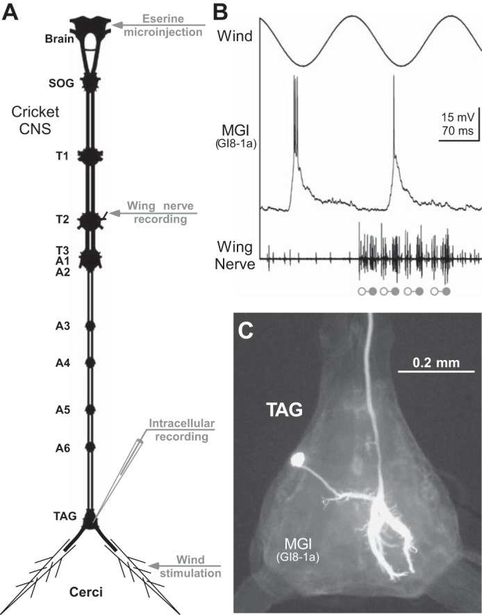
Experimental design. A: diagram of the cricket central nervous system (CNS) indicates the location of the mesothoracic wing-nerve (T2-N3A) recording, eserine injection into the brain, intracellular recording in the terminal abdominal ganglion (TAG), and the cercal wind stimulation. B: synaptic and spike response of a wind-sensitive cercal giant interneuron (middle trace) to sinusoidal cercal wind stimulation (top trace) during fictive singing. Open and solid circles indicate opener and closer motoneuron spike bursts, respectively, of a four-syllable chirp in the wing nerve recording (bottom trace). C: photomicrograph of the Lucifer Yellow labeled interneuron in the TAG allows subsequent anatomical identification as the median giant interneuron (MGI), also known as GI8-1a, according the classification by Jacobs and Murphey (1987).