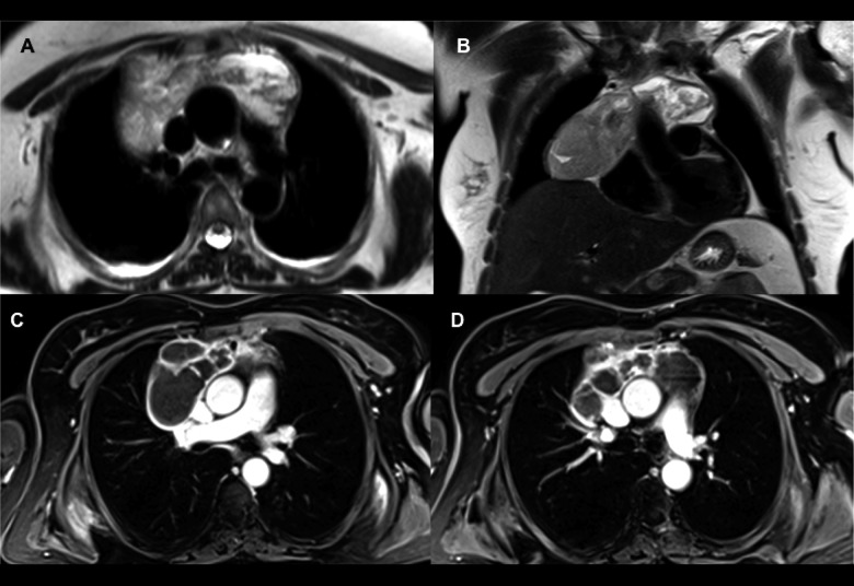
Figure 2.
T2 TSE axial (A) and coronal (B) MR images: the mass is hyperintense compared to muscular tissues but presents multiple internal septa. T1 VIBE axial MR images after after intravenous contrast agent administration (C, D): the mass shows clear internal fluid content with a significant enhancement of walls and septa, particularly in the anterior portion.

