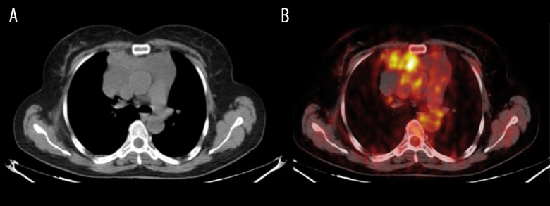
Figure 3.

(A, B) Axial 18F-FDG PET/CT shows tracer uptake in the capsular portion of the mass (SUV max 2) and focal intense uptake (SUV max 5.7) was clearly detected in the middle of the lesion.

(A, B) Axial 18F-FDG PET/CT shows tracer uptake in the capsular portion of the mass (SUV max 2) and focal intense uptake (SUV max 5.7) was clearly detected in the middle of the lesion.