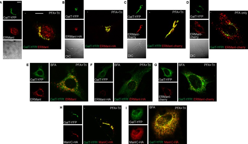
FIGURE 4:
ERManI localization is altered in fixed and permeabilized cells. (A–C) NIH 3T3 cells transfected with GalT-YFP only (A) or together with ERManI-HA (B) or ERManI-cherry (C) were fixed with 3% PFA and permeabilized with 0.5% Triton. They were then stained with mouse anti-HA (B) or mouse anti-ERManI (A; for endogenous ERManI), followed by goat anti-mouse Dylight 594 or left unstained (C). There is colocalization between all versions of ERManI and the Golgi marker. Bars, 10 μm. (D) When fixed with 3% PFA but left unpermeabilized, cells show ERManI-cherry in vesicles, distinct from GalT-YFP. (E–G) Cells treated with 5 μg/ml BFA for 1 h show an expected redistribution of the Golgi marker GalT-YFP to an ER pattern, whereas endogenous ERManI (E), ERManI-HA (F), and ERManI-cherry (G) are all in a vesicular pattern. Staining as in A–C. (H, I) Cells expressing GalT-YFP and the Golgi mannosidase Man1C-HA were either left untreated (H) or treated with BFA (I). They were then fixed as in A. Both proteins colocalize before (Golgi pattern) and after BFA treatment (ER pattern).

