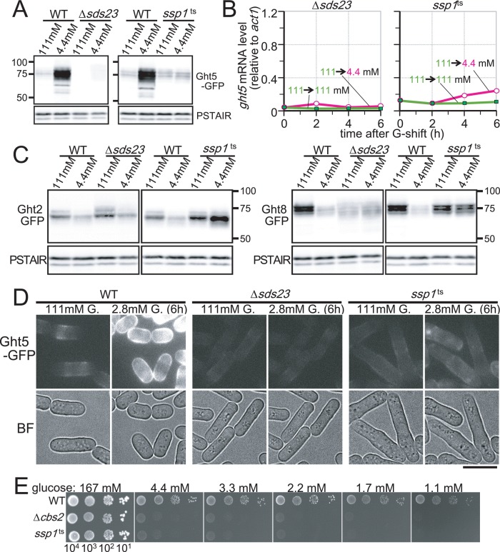
FIGURE 6:
Sds23 and Ssp1 are required for elevated expression of Ght5 under low glucose. (A) Immunoblotting for Ght5-GFP in WT, Δsds23, and ssp1-837 (ssp1ts) mutant strains. WT and mutant cells were cultivated in EMM2 containing either 111 or 4.4 mM glucose for 6 h at 33°C before sample preparation. Blots with anti-PSTAIR antibody are shown as the loading control. (B) Time course of ght5+ mRNA levels was examined in the Δsds23 and the ssp1ts strains. Exponentially growing cells were transferred to fresh EMM2 containing 111 (filled rectangles) or 4.4 mM (open circles) glucose and cultivated at 33°C. mRNA levels of ght5+ relative to those of act1+ were measured by RT-qPCR at 2-h intervals. (C) Immunoblotting for Ght2- and Ght8-GFP in WT, Δsds23, and ssp1-837 (ssp1ts) mutant strains. WT and mutant cells were cultivated in EMM2 containing either 111 or 4.4 mM glucose for 6 h at 33°C before sample preparation. Blots with anti-PSTAIR antibody are shown as the loading control. (D) Intracellular localization of Ght5 was examined in Δsds23 and ssp1ts mutants, as well as in WT control. Cells harboring Ght5-GFP were cultivated at 33°C in a microfluidic perfusion chamber with a continuous supply of medium, and the glucose concentration was switched from 111 to 2.8 mM. GFP and brightfield (BF) microscopy images of the cells before medium switching and after 6-h cultivation in 2.8 mM glucose are shown. All GFP images here were processed under the same conditions. Bar, 10 μm. (E) Aliquots of 104 cells of the WT, Δcbs2, and ts ssp1-837 mutant strains were serially diluted 10-fold, spotted onto YES medium plates containing the indicated concentrations of glucose, and incubated at 33°C for 3 d. See also Supplemental Figure S3.

