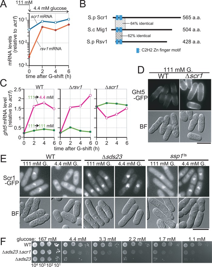
FIGURE 7:
Nuclear exclusion of Scr1, which represses transcription of ght5+ in high glucose, requires the presence of Sds23 and Ssp1. (A) Levels of rsv1+ (filled diamonds) and scr1+ (open squares) mRNAs relative to that of act1+ were determined by RT-qPCR in the cells cultivated in 4.4 mM glucose medium for 0, 2, 4, and 6 h after the shift from 111 mM glucose. (B) S. pombe Scr1 and Rsv1 proteins are schematically presented together with their S. cerevisiae homologue, Mig1p. The length of each protein is indicated at the right. Although Scr1 and Rsv1 are the closest fission yeast homologues to Mig1p, their sequence similarities, which are represented as percentage identity to Mig1p, are limited to small regions surrounding C2H2-type zinc finger motifs, whose positions are indicated by the filled boxes. (C) Time course of ght5+ mRNA levels was examined in WT, Δrsv1, and Δscr1 strains. Exponentially growing cells were transferred to fresh EMM2 containing 111 (filled rectangles) or 4.4 mM (open circles) glucose and cultivated at 33°C. The mRNA levels of ght5+ relative to those of act1+ were measured by RT-qPCR at 2-h intervals. (D) Micrographs of the WT and the Δscr1 cells expressing Ght5-GFP under the native promoter. Exponentially growing cells in EMM2 containing 111 mM glucose at 33°C were harvested by centrifugation and resuspended in a small amount of the same medium. Ght5-GFP fluorescence and brightfield (BF) microscopy images showing cell shape were obtained immediately without fixation. Fluorescence images here were processed under the same conditions. Bar, 10 μm. (E) Intracellular localization of Scr1-GFP in WT, Δsds23, and ssp1-537 cells. Cells expressing Scr1-GFP were transferred from EMM2 with 111 mM glucose to EMM2 with either 111 or 4.4 mM glucose and cultivated for 4 h before sampling. Scr1-GFP fluorescence and BF microscopy images were obtained immediately without fixation. Bar, 10 μm. (F) Aliquots of 104 cells of WT, Δsds23 Δscr1 double-mutant, and Δsds23 mutant strains were serially diluted 10-fold, spotted onto YES medium plates containing the indicated concentrations of glucose, and incubated at 30°C for 3 d. See also Supplemental Figure S4.

