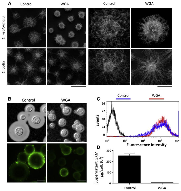
Fig. 6.
Phenotypic analysis of capsule formation in C. neoformans after exposure of the fungus to WGA at 500 μg/ml. Scanning electron microscopy (A) demonstrated that capsular dimensions of C. neoformans, but not of C. gattii, were affected by treatment with WGA. Capsule morphology is shown for samples of C. neoformans and C. gattii populations (left panels; scale bar, 5 μm) and for individual cells (right panels; scale bar, 2 μm). SEM observations of C. neoformans correlate with the phenotype observed by India ink counter staining (B, upper panels) and with the regular recognition of surface polysaccharides by a monoclonal antibody to GXM, as demonstrated by immunofluorescence microcopy (B, lower panels) and flow cytometry (C). Under this condition, the concentration of extracellular GXM is significantly smaller in culture supernatants of WGA-treated cells (D, P < 0.00001).

