Fig. 4.

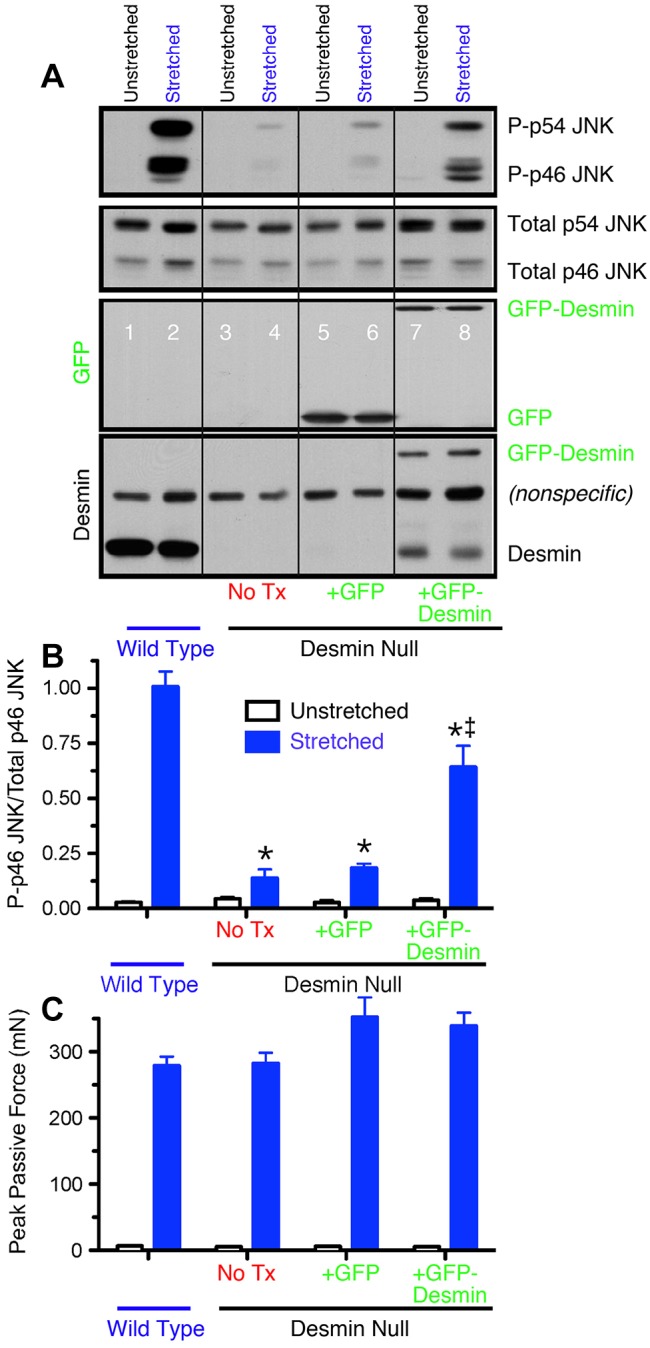
GFP–desmin transfection rescues the normal JNK signaling response. (A) Western blots of p46 JNK and p54 JNK phosphorylation in stretched and unstretched extensor digitorum longus muscles. Antibodies used are, from top to bottom, phosphorylated JNK, total JNK, GFP and desmin. (B) Quantification of p46 JNK western blots, such as those shown in A. Note the strong phosphorylation of p46 JNK and p54 JNK upon stretch in WT muscles and desmin-null muscles transfected with GFP–desmin but not in untreated desmin-null muscles or desmin-null muscles transfected with GFP alone. Data shown are mean±s.e.m. (n = 4 for each experiment). Also note that p46 JNK phosphorylation is partially rescued in desmin-null muscles transfected with GFP–desmin. Identical results were obtained for p54 JNK (data no shown). (C) Peak mechanical force reached during cyclic deformation of different treatment groups. No significant difference was observed between WT and desmin-null groups for any treatment demonstrating that the signaling difference was not simply a secondary effect of altered cellular mechanics. Data shown are mean±s.e.m. (n = 4 for each experiment).
