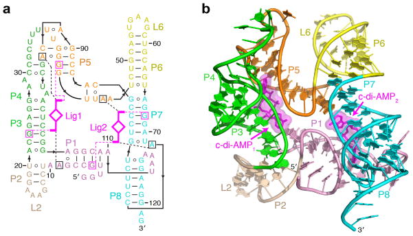
Figure 1. Overall structure and schematics of the T. pseudethanolicus c-di-AMP riboswitch.
(a) Secondary structure schematic of the riboswitch fold observed in the crystal structure of the complex. Key base-specific hydrogen bonding and stacking interactions between ligand (magenta) and RNA nucleotides (squared) are shown as dashed magenta and black lines, respectively. (b) Overall c-di-AMP riboswitch structure in a ribbon representation showing front view. Ligands are in stick and surface representations.

