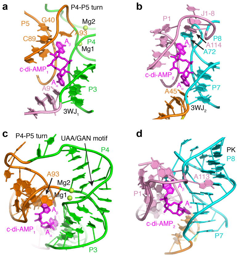
Figure 2. Structural elements of the T. pseudethanolicus c-di-AMP riboswitch.
(a, b) Structures of the ligand binding sites 1 (a) and 2 (b) shown with nucleotides that either interact with ligands directly or depend on ligand binding. (c) Structure of the double turn bound to c-di-AMP. Mg2+ cations are shown as green spheres. (d) Structure of the pseudoknot and J1-8 region bound to c-di-AMP.

