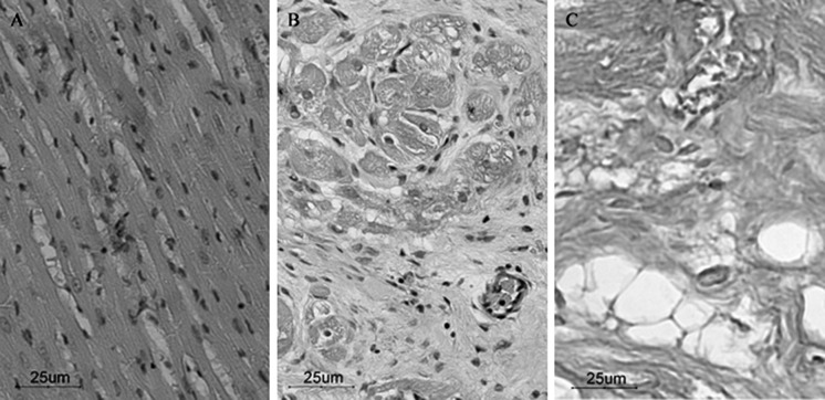
Fig. 2.
HE staining. a Normal myocardial cells are arranged in neat rows and have normal cell size and morphology; b infarcted area of the junction with normal myocardium, and remnants of infarct border myocardial tissue can be seen; c infarct zone has mainly scar formation, with a large number of collagen fibers and focal steatosis (inverted phase contrast microscope, ×200)

