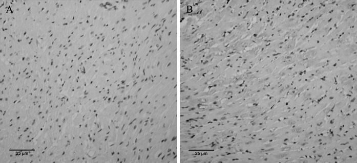
Fig. 4.

Immunohistochemical staining results of VEGF expression at 4 weeks after cell transplantation. The brown areas denote VEGF positive expression. a PBS injection at 7 days after MI, b MSCs + G-CSF injection at 7 days after MI (inverted phase contrast microscope, ×200)
