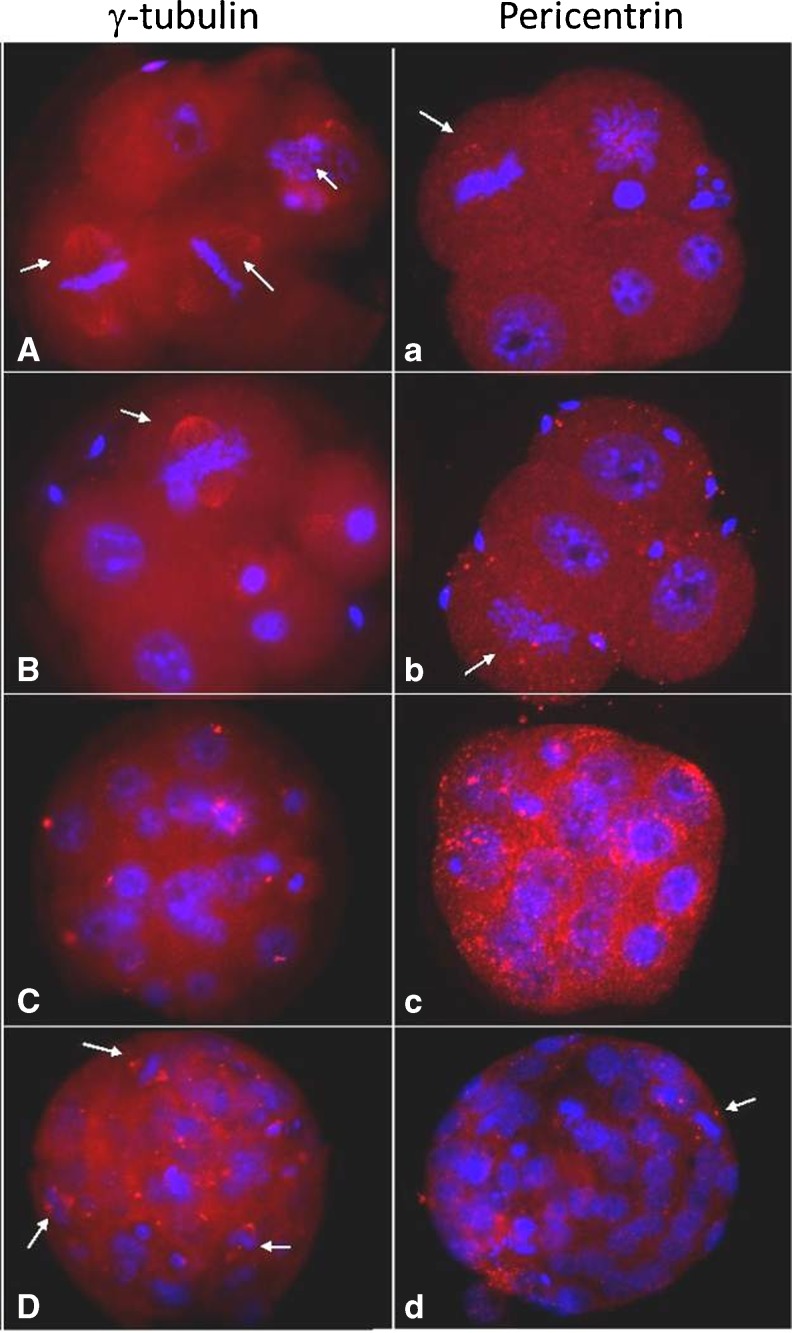
Fig. 5.
γ-tubulin (A–D) and pericentrin (a–d) pattern in mouse embryos. Chromatin is shown in blue (Hoechst staining). Note distribution of γ-tubulin (A, B), not pericentrin (a, b), forming the spindle during the first cleavages. γ-tubulin exhibits a punctuate perinuclear localization at compacted morula (C) and forms the spindle proper in blastocysts stage embryos (D). Pericentrin exhibits an apical distribution in 8-cell stage embryos (c) and forms the spindle poles in the blastocyst stage (d). Arrow indicate cell in metaphase

