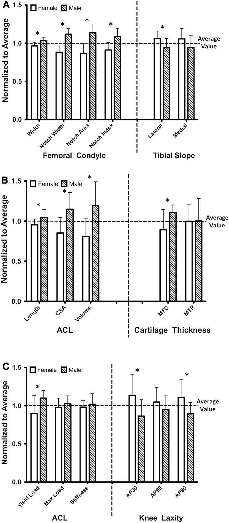
Fig. 4A–C.
Measurements for each sex were normalized to the pooled male and female values (mean ± SD) to better characterize the role of the sex on quantified (A) bony geometry, (B) ACL and cartilage geometry, and (C) structural variables. Variables that are different (p < 0.05) between the males and females are indicated by an asterisk. CSA = cross-sectional area; MFC = medial femoral condyle; MTP = medial tibial plateau.

